Treating Founder (Chronic Laminitis) without Horseshoes
Understanding X-rays
(Full-sized photos--better for printouts.)
I have had the advantage of working in the black and white
film department of a custom photo lab for a couple of years, and this has given me
insights into x-rays that many people do not have.
Many horse owners are under the impression that x-rays are consistent, and that one can
fairly compare x-rays taken at different times. This is similar to the illusion
that many people have that their pictures did not "turn out" if the first lab
they take their negatives to for prints makes some bad-looking prints. Those of us with
darkroom experience know this is not so at all. The same negative can be printed hundreds
of different ways--all different light/dark variations, degrees of contrast, color
balance, etc.
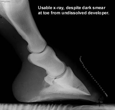
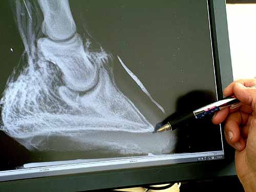
The x-ray below has a lot going for it. The hoof is up on a board, with the film holder resting on the ground, so the edges of the film holder will not obscure the bottom of the foot, but actually show it all. It has enough exposure to see detail, and enough contrast. I taped a beaded key chain down the center front of the toe so I can see exactly where the toe profile is. If the exposure is so light that you don't need a metal marker on the toe to show where the toe profile is, then you won't be able to see very well if the bottom of the coffin bone is ground-parallel. Another plus with using a beaded chain instead of a nail or piece of wire is that it follows the contours of the hoof exactly, showing dishing accurately. The x-ray machine was resting on the ground, its beam aimed ground-parallel--another important point. If the machine is aimed downwards, for instance, you will not be able to see very well if the coffin bone is ground-parallel. There was one little black smear on the bottom of toe, indicating some undissolved developer solution crystals, but overall, it is a usable and readable x-ray.
What does the fact that x-rays can vary
widely in quality mean to you, a horseowner?
It means that if between one set of x-rays and another, if it seems your horse's coffin
bone is eroding....maybe it isn't. Maybe the later x-ray had a higher overall
density that made the coffin bone LOOK smaller than it would look on a less dense x-ray.
This is obvious to people with darkroom backgrounds. We would be the first to want to do
densitometer readings all over both x-rays to determine degree of contrast and overall
exposure. But the horse owner with no darkroom background might be in a state of
panic, thinking the coffin bone is dissolving, and could even make horrible decisions,
like euthanasia, when it may in fact not be as bad as it looks.
X-rays are really just black and white negatives that are exposed by x-ray radiation
rather than light. The principles are the same, though.
The developer phase is quite critical. Temperature fluctuations of even 1 degree F. can
very strongly impact the overall density and contrast range of the final results.
Factors that result in weak, uncontrasty negatives and low density:
1. Depleted, exhausted developer.
2. Developer too cold for the time the negatives are "in the soup."
3. Under-exposure by the x-ray machine. Distances between the x-ray machine and the hoof
are critical; the machine being even a little further away will result in a "Light
fall-off" effect similar to flash photography.
4. Inadequate agitation during the developer phase of processing.
5. Exposures have to be increased for bigger, denser feet. My mule's feet turned out to be
weaker exposures than my horse's for this reason.
6. Outdated x-ray film can have an overall
dichroic fog, lowering overall contrast. Film that is not outdated can still age
prematurely if it not kept cool.
Factors that result in dense, overly contrasty negatives are the opposite of the
above. In addition, old developer that has been over-replenished with developer
replenisher results in too contrasty a result. Replenisher is a cheap measure when
it really is time to dump the developer and start over.
Other unbelievably sloppy problems shown in these images:
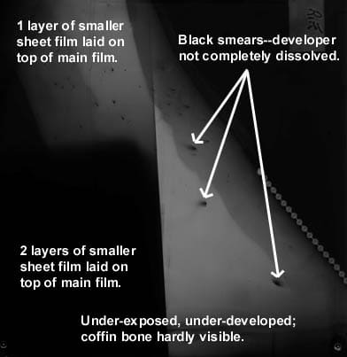
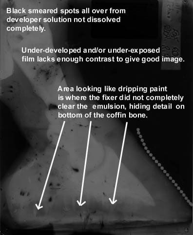
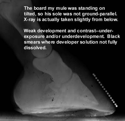
1. Dark smeared spots, the result of developer crystals not being completely dissolved.
Developers usually need to be mixed with the water temperature at least 90 degrees
F.....and then used, usually, at around 70 degrees F.
2. One image shows there were 2 smaller pieces
of film in one film holder, obscuring the main image....see above.
3. One image is of a tilted hoof; it is impossible to get an accurate reading on whether
or not the coffin bone is truly ground-parallel unless the hoof is ground-parallel on a
board, and the x-ray machine is aimed ground-parallel, instead of looking up or down at
the hoof.
4. One image shows that the emulsion was not
completely cleared by the fixer bath--looks like translucent, dripping paint, obscuring
the bottom of the coffin bone. This can be caused by depleted fixer, fluid level in the
fix tank too low to cover the film in its hanger, and inadequate agitation. See
above.
5. Three of the images are weak and uncontrasty due to inadequate development and/or
depleted developer.
Hand-developed x-rays in vets' offices, usually done by receptionists with no darkroom
background, are more likely to have problems. Even x-rays developed on automatic machines
are not consistent, however. I worked with a Kodak Versamat. Even slight changes
in the wash water temperature had
striking results on negative contrast and overall exposure. We ran test strips daily. Test
strips are a step wedge scale, very consistently exposed by Kodak. We pulled them out of
the fridge daily, and read them on the densitometer after they were processed. The
variations in the readings were unsettling, to say the least.
So--please bear in mind that x-rays have to be looked at critically. I am
intrigued with fluoroscopes because at least the darkroom variables are eliminated,
yielding more fairly comparable, repeatable results. They can have the added benefit of
creating immediate digital images that can be saved onto floppy disks, emailed, etc. Their
cost is prohibitive, though. I also understand that they do not render detail as
fine as a conventional x-ray. The machine I had heard about for doing hooves did not
cover as large an area, either--not the full 8" x 10" area I would want to see.
Ideal x-ray technique--Ric Redden, DVM taking digital x-rays and venograms at the Laminitis Conference, Nov. 2005:
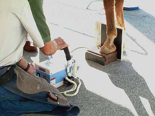
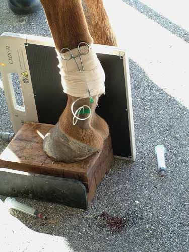

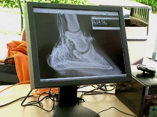
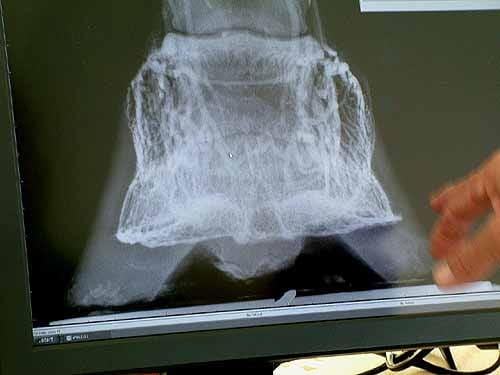
You want to stand both fores, or both hinds, up on wooden blocks. Otherwise, the bottom of the film holder will obscure some important information, like how thick the soles are and how much concavity you really have. The x-ray machine should be on the ground, aimed horizontally. If the x-ray machine is held higher up and is looking somewhat down, this will skew your results. It should also be directly out to the side, not at some other angle. To mark the center of the toe, the ideal solution is radio-opaque paste. This could also be used to mark the apex of the frog. Lacking that, you can tape a short piece of beaded key chain down the center front of the toe so you can see exactly where the toe profile is on the x-ray. Otherwise, it will blend into the black background, and you won't be able to see and you won't be able to see truly where the leading edge of the toe wall is. Taping on a nail or piece of wire is not as good as beaded key chain because these don't follow the toe wall form as exactly. The radio opaque paste is even better, and much quicker to apply, but your vet may not have it. Also, many people use a thumb tack to mark the apex of the frog. Also, it is important to have the x-ray plate as close to the hoof as possible for better detail.
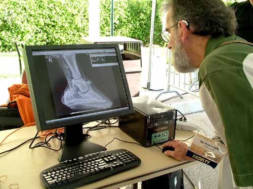
These photos show how Ric Redden, DVM took x-rays at the laminitis conference recently, which incorporates all the points I am talking about here. He had an awesome digital unit that gave you instant results on a computer monitor, and also allowed you to zoom in on details. The digital x-rays are instant, and can be saved to CDs or emailed to clients, and are available immediately to a farrier working on the case. If your exposure is poor, you will know this instantly, unlike with film-based x-rays. It eliminates dark room quality control problems as well. The photos were taken of Redden doing a venogram. This involves injecting contrast dye into the foot, and getting an image of not only the bones, but also of the circulation in the foot. When circulation is compromised, it will show up on the venogram, during acute laminitis. When circulation improves, that, too, will show up on a venogram. Venograms could potentially give your vet a good idea of how badly circulation is affected during acute laminitis, and could also help in predicting early on how large an area will have damage, or how severe a case you will be facing. Another interesting aspect of venograms is that they seem to temporarily reduce pain.
For more information on how venograms are
taken:
http://www.nanric.com/venogram_technique.html
Readers! If you have any bad x-rays you would like to submit to this page for inclusion, please contact me. I would be happy to scan your x-rays and return them to you.
Mail x-rays to me at:
Gretchen Fathauer
PO Box 307
Duncan Falls, OH 43734
Phone: 740-674-4492
gretchenfathauer@prodigy.net
Back to home page--Table of Contents
Article in sections with "thumbnail" photos
for fastest
downloads:
1
9
17
2
10
18
3
11
19
4
12
20
5
13
21
6
14
22
7
15
23
8
16
24
NAVICULAR
Article in sections with full-sized photos
for print-outs:
1
9
17
2
10
18
3
11
19
4
12
20
5
13
21
6
14
22
7
15
23
8
16
24
NAVICULAR
To
Strasser case studies--thumbnail photos for faster
downloads
To
Strasser case studies--large photos
Please sign my guest
book!
Photos of my pets
My farm
Share Barefoot
success stories on this page
Buy or sell used HORSE
BOOTS Natural
board Barn
Listings

Click here to subscribe to naturalhorsetrim
(I strictly moderate this listserv to weed out "fluff.")
Send Email to Gretchen Fathauer, or call (740) 674-4492
Copyright by Gretchen Fathauer, 2013. All rights reserved.