Treating Founder (Chronic Laminitis) without Horseshoes, Section 9
Faster-loading version with "thumbnail" photos
(Click on small photos to see larger versions)
WHY DO LAMINAE MAINLY DIE OFF IN THE TOE AREA?
Not having the bottom of the coffin bone ground-parallel (heels too high) overstresses the toe laminae and also sets up inflammation.
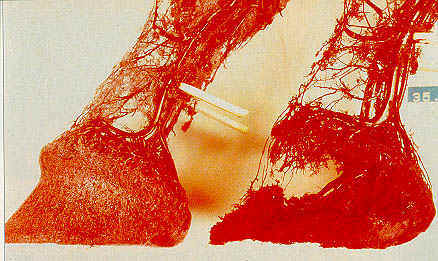
Below is an image of 2 "corrosion castings." Cool-setting polymers are injected into the blood vessels. After tissue is removed, the remaining casting gives a clear picture of how circulation is changed during laminitis.
Circulatory Differences--Normal Foot and Foot in Acute Laminitis
Normal Foot, left--Foot in Acute Stage of Laminitis, right
(Photo of Dr. Christopher Pollitt's corrosions, courtesy of American Farriers Journal)
NOTICE that the coronet bands are sloped more and
the heels are lower and not under-run in the more normal foot.
Pollitt's corrosions show that the normal foot has uniform circulation, while in a laminitic foot, there is a total lack of circulation in the area in front of the coffin bone. This is why you end up having enough laminae destroyed to result in rotation. Blood is engorged in the heels and pasterns and other soft areas that can expand to accommodate swelling.
These circulatory blockages Pollittt's corrosions show in the toe raise the horse's blood pressure. As long as you are feeling a bounding pulse in the horse's blood vessels at the rear of the pasterns, you can tell the blood pressure is still elevated due to resistance to circulation in the foot...a bad sign! Still in the acute stage of laminitis!
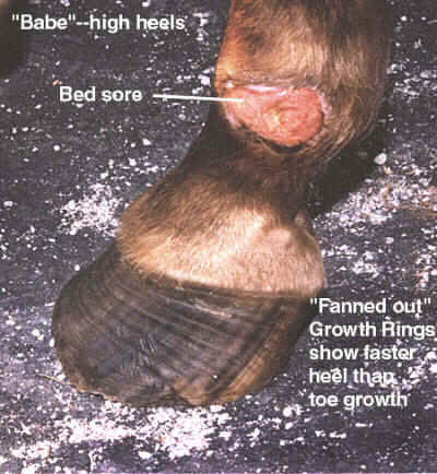
"Babe," Sept./Oct. '98
How Babe's high heels contribute to slower growth in toes than heels: high heels put extra weight on the toe, compressing the coronary corium by leveraging into it, which reduces circulation in the toe area of the coronet. Also, lack of weight on the heels makes them grow faster, and more weight on the toes make them grow slower.
A more far-reaching consequence of poor circulation and resultant reduced hoof growth that Dr. Strasser talks about in her book: poor hoof circulation, leading to slower hoof growth, results in a back-up of protein in the body, protein that should have been utilized for growing hooves faster. This excess protein stresses the skin, liver and kidneys, resulting in skin problems (eczema and slow shedding out) and metabolic disturbances. Some of this sounds similar in some respects to Cushing's Disease.
The solution to excess protein building up in the blood is not to just put your horse on low-protein or starvation diet. The solution is to get enough movement and hoof mechanism so the horse actually can put the protein in his diet to good use. I would also like to note that Linsey McLean's grain formula for foundered, low thyroid and Cushings horses is high-protein, but lower in ingredients with a high glycemic index, such as sugary, simple carbohydrates. More info: http://www.vitaroyal.com/FounderAllergy.html There may be some similarities between some of these conditions and sugar metabolism problems in humans, such as Diabetes type II. I have had my guys on Linsey's high-protein mix for some time, and feeding them more of it than I used to feed of other grains in the past. It has not foundered any of them, or made them hyper, but it has added some muscle.
The recipe for Linsey's high-protein non-grain feed mix:
Linseed (flax) meal or pellets 300 lbs
soybean meal ---150 lbs
bran (preferably wheat) ---75 lbs
calcium carbonate (feed grade only) ---15 lbs (Delete if you are in an area with a lot of limestone in the water supply)
magnesium oxide (feed grade only)---12 lbs
high iodine trace mineral salt (commonly used for foot rot in cattle, not just regular TM salt) such as Morton's TM+4 ---6 lbs
(Linsey cautions you that this formula will probably need some vitamin and mineral supplementation.)
There is disagreement on what causes the loss of circulation within the laminae:
Dr. Pollitt's view, (See Equus Magazine, April '92), is that the lack of circulation is caused by AVA's (arteriovenous anastomoses) remaining dilated for hours. Normally, these pathways between the veins and arteries, which shunt blood away from the nutritive capillary bed of the laminae when open, remain closed. They normally only open briefly and repeatedly in situations like the horse standing in snow or icy water, to prevent chilling of the foot. The hope is that if the AVA's paralysis can be better understood, perhaps we can prevent their remaining frozen open during an attack of laminitis by either neuroactive drugs, immunologic or biomolecular approaches. Research is still ongoing.
David Frederick, DVM's view, (See American Farriers Journal, September/October '97) is that the lack of circulation results from the swelling, inflammation and edema within the insensitive laminae. He believes this swelling squeezes the blood vessels shut. He draws this conclusion from Dr. Pollitt's microscopic studies of both normal and foundered laminae. The insensitive laminae appear to be bloated with edema and water, thereby compressing the smallest blood vessels in the sensitive laminae. This cuts off circulation.
Dr. Frederick's view, implicating hydraulic pressure inside the laminae, is closer to Dr. Strasser's view, but she believes that a lack of hoof mechanism due to incorrect hoof form and inactivity are the real culprits. Jaime Jackson was hard-pressed to find foundered wild horses freshly mustered in from the range. These wild horses constantly maintained a short toe, low heel foot shape, and walked 15 miles a day on average, meaning that they had excellent hoof mechanism. Such a horse exposed to a laminitis trigger is better able to weather it as a minor episode that passes rather uneventfully in a few days without rotation.
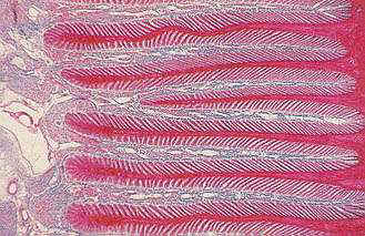
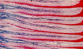
Top, Normal laminae. Red and pink are the insensitive laminae; blue sensitive.
Bottom, Acute laminitis. More white, which Dr. Frederick says is water, or edema.
Sabine Kells describes this as wound secretion, a product of inflammation.
This specimen is only a few hours into laminitis, prior to any laminae separating.
(Dr. Christopher Pollitt's microscopic images courtesy of American Farriers Journal.)
Back to home page--Table of Contents
Article in sections with "thumbnail" photos for fastest downloads:
1 9 17
2 10 18
3 11 19
4 12 20
5 13 21
6 14 22
7 15 23
8 16 24
NAVICULAR
Article in sections with full-sized photos for print-outs:
1 9 17
2 10 18
3 11 19
4 12 20
5 13 21
6 14 22
7 15 23
8 16 24
NAVICULAR
To Strasser case studies--thumbnail photos for faster downloads
To Strasser case studies--large photos
Please sign my guest book! Photos of my pets My farm
Share Barefoot success stories on this page
Buy or sell used HORSE BOOTS Natural board Barn Listings
Click here to subscribe to naturalhorsetrim
(I moderate this listserv to weed out "fluff.")
Send Email to Gretchen Fathauer, or call (740) 674-4492
Copyright by Gretchen Fathauer, 2015. All rights reserved.